Posts

Showing posts from January, 2022

Types of Neurotransmitters

Image
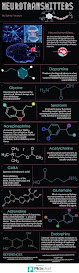
  Types of Neurotransmitters Neurotransmitters, at the highest level, can be sorted into two types: small-molecule transmitters and neuropeptides. Small-molecule transmitters, like dopamine and glutamate, typically act directly on neighboring cells. The neuropeptides, small molecules like insulin and oxytocin, work more subtly, modulating, or adjusting, how cells communicate at the synapse. To date, scientists have identified more than 60 distinct types of neurotransmitters in the human brain, and most experts say there are more left to discover. These powerful neurochemicals are at the center of neurotransmission, and, as such, are critical to human cognition and behavior. Acetylcholine Acetylcholine (Ach) was the first neurotransmitter discovered. It is a direct action small-molecule that works primarily in muscles, helping to translate our intentions to move into actual actions as signals are passed from the neurons into the muscle fiber. But it also has other roles in the brain, in

The HIV Life Cycle

Image
  The HIV Life Cycle In order for viruses to reproduce, they must infect cells in the body. Viruses are not technically alive: They are like a brain without a body. So in order to make more copies of itself, a virus must hijack our cells and use them to make new viruses. But how does that happen? Your body constantly makes new skin and blood cells, and each cell often makes new proteins in order to stay alive and reproduce. Viruses hide their own DNA in the DNA of the cell. So when the cell tries to make its own proteins, it accidentally makes new viruses as well. HIV can infect many types of cells in the body, but it mostly infects cells in the immune system. Once infected, a cell can produce hundreds of new copies of HIV. Several kinds of immune cells have proteins on their surface, called CD4 receptors. HIV searches for these because the CD4 protein helps the virus bind to the cell. The main target for HIV is a white blood cell called a T4 lymphocyte, or “T helper cell”. The T4 cell

FLOW OF CSF

Image
  FLOW OF CSF Cerebrospinal fluid is constantly produced at a secretion rate of 0.2-0.7 ml/min, meaning that there is 600–700 ml of newly produced CSF per day. Since the total volume of CSF averages around 150-270 mL, this means that the entire volume of CSF is replaced around 4 times per day. The pathway of the cerebrospinal fluid is as follows: The CSF passes from the lateral ventricles to the third ventricle through the interventricular foramen (of Monro). From the third ventricle, the CSF flows through the cerebral aqueduct (of Sylvius) to the fourth ventricle. From the fourth ventricle, some CSF flows through a narrow passage called the obex and enters the central canal of the spinal cord. However, the majority of CSF passes through the apertures of the fourth ventricle; the median aperture (of Magendie) and two lateral apertures (of Luschka). Via these openings, the CSF enters the cisterna magna and cerebellopontine cisterns, respectively. From there, the CSF flows through the s

Types of pain

Image
  Types of pain Acute pain Acute pain means the pain is short in duration (relatively speaking), lasting from minutes to about three months (sometimes up to six months). Acute pain also tends to be related to a soft-tissue injury or a temporary illness, so it typically subsides after the injury heals or the illness subsides. Acute pain from an injury may evolve into chronic pain if the injury doesn’t heal correctly or if the pain signals malfunction. Chronic pain Chronic pain is longer in duration. It can be constant or intermittent. For example, headaches can be considered chronic pain when they continue over many months or years – even if the pain isn’t always present. Chronic pain is often due to a health condition, like arthritis, fibromyalgia, or a spine condition. Neuropathic pain Neuropathic pain is due to damage to the nerves or other parts of the nervous system. It is often described as shooting, stabbing, or burning pain, or it feels like pins and needles. It can also affect

CARDIAC CONDUCTION SYSTEM

Image
  CARDIAC CONDUCTION SYSTEM Step 1: Pacemaker Impulse Generation The first step of cardiac conduction is impulse generation. The sinoatrial (SA) node (also referred to as the pacemaker of the heart) contracts, generating nerve impulses that travel throughout the heart wall. This causes both atria to contract. The SA node is located in the upper wall of the right atrium. It is composed of nodal tissue that has characteristics of both muscle and nervous tissue. Step 2: AV Node Impulse Conduction The atrioventricular (AV) node lies on the right side of the partition that divides the atria, near the bottom of the right atrium. When the impulses from the SA node reach the AV node, they are delayed for about a tenth of a second. This delay allows atria to contract and empty their contents into the ventricles prior to ventricle contraction. Step 3: AV Bundle Impulse Conduction The impulses are then sent down the atrioventricular bundle. This bundle of fibers branches off into two bundles and

THE LIFE CYCLE FOR MALARIA

Image
  THE LIFE CYCLE FOR MALARIA IN MAN 1. Malaria infection begins when an infected female Anopheles mosquito bites a person, injecting Plasmodium parasites, in the form of sporozoites, into the bloodstream. 2. The sporozoites pass quickly into the human liver. 3.The sporozoites multiply asexually in the liver cells over the next 7 to 10 days, causing no symptoms. 4.In an animal model, the parasites, in the form of merozoites, are released from the liver cells in vesicles, journey through the heart, and arrive in the lungs, where they settle within lung capillaries. The vesicles eventually disintegrate, freeing the merozoites to enter the blood phase of their development.* 5.In the bloodstream, the merozoites invade red blood cells (erythrocytes) and multiply again until the cells burst. Then they invade more erythrocytes. This cycle is repeated, causing fever each time parasites break free and invade blood cells. 6.Some of the infected blood cells leave the cycle of asexual multiplicati

MECHANISM OF SWALLOWING

Image
  MECHANISM OF SWALLOWING 1. The Pre-oral Phase – Starts with the anticipation of food being introduced into the mouth – Salivation is triggered by the sight and smell of food (as well as hunger) 2. The Oral Phase – The lips close and form a seal – Chewing / mastication of food begins – The food is mixed with saliva to form a bolus – The bolus is then transferred to the back of the mouth 3. The Pharyngeal Phase – Reflexive (reflex response- i.e. not planned) initiation of the swallow. – The nasal cavity is sealed when the soft palate raises (that prevents food/fluid coming out of the nose) – The larynx (voice box) moves upwards and forward – The vocal folds close, and then the epiglottis closes over the airway (breathing stops momentarily) – The pharynx pushes the bolus down (by contracting in a stripping motion) – The upper oesophageal sphincter opens to allow the bolus through – The upper oesophageal sphincter closes after the bolus has passed through in order to prevent the bolus m

𝙁𝙀𝙍𝙏𝙄𝙇𝙄𝙕𝘼𝙏𝙄𝙊𝙉

Image
  𝙁𝙀𝙍𝙏𝙄𝙇𝙄𝙕𝘼𝙏𝙄𝙊𝙉   𝘏𝘶𝘮𝘢𝘯 𝘧𝘦𝘳𝘵𝘪𝘭𝘪𝘻𝘢𝘵𝘪𝘰𝘯 𝘪𝘴 𝘵𝘩𝘦 𝘶𝘯𝘪𝘰𝘯 𝘰𝘧 𝘢 𝘩𝘶𝘮𝘢𝘯 𝘦𝘨𝘨 𝘢𝘯𝘥 𝘴𝘱𝘦𝘳𝘮 , 𝘶𝘴𝘶𝘢𝘭𝘭𝘺 𝘰𝘤𝘤𝘶𝘳𝘳𝘪𝘯𝘨 𝘪𝘯 𝘵𝘩𝘦 𝘢𝘮𝘱𝘶𝘭𝘭𝘢 𝘰𝘧 𝘵𝘩𝘦 𝘧𝘢𝘭𝘭𝘰𝘱𝘪𝘢𝘯 𝘵𝘶𝘣𝘦 . 𝘛𝘩𝘦 𝘳𝘦𝘴𝘶𝘭𝘵 𝘰𝘧 𝘵𝘩𝘪𝘴 𝘶𝘯𝘪𝘰𝘯 𝘪𝘴 𝘵𝘩𝘦 𝘱𝘳𝘰𝘥𝘶𝘤𝘵𝘪𝘰𝘯 𝘰𝘧 𝘢 𝘻𝘺𝘨𝘰𝘵𝘦 𝘤𝘦𝘭𝘭 , 𝘰𝘳 𝘧𝘦𝘳𝘵𝘪𝘭𝘪𝘻𝘦𝘥 𝘦𝘨𝘨 , 𝘪𝘯𝘪𝘵𝘪𝘢𝘵𝘪𝘯𝘨 𝘱𝘳𝘦𝘯𝘢𝘵𝘢𝘭 𝘥𝘦𝘷𝘦𝘭𝘰𝘱𝘮𝘦𝘯𝘵 . 𝘚𝘤𝘪𝘦𝘯𝘵𝘪𝘴𝘵𝘴 𝘥𝘪𝘴𝘤𝘰𝘷𝘦𝘳𝘦𝘥 𝘵𝘩𝘦 𝘥𝘺𝘯𝘢𝘮𝘪𝘤𝘴 𝘰𝘧 𝘩𝘶𝘮𝘢𝘯 𝘧𝘦𝘳𝘵𝘪𝘭𝘪𝘻𝘢𝘵𝘪𝘰𝘯 𝘪𝘯 𝘵𝘩𝘦 𝘯𝘪𝘯𝘦𝘵𝘦𝘦𝘯𝘵𝘩 𝘤𝘦𝘯𝘵𝘶𝘳𝘺 .     𝘛𝘩𝘦 𝘱𝘳𝘰𝘤𝘦𝘴𝘴 𝘰𝘧 𝘧𝘦𝘳𝘵𝘪𝘭𝘪𝘻𝘢𝘵𝘪𝘰𝘯 𝘪𝘯𝘷𝘰𝘭𝘷𝘦𝘴 𝘢 𝘴𝘱𝘦𝘳𝘮 𝘧𝘶𝘴𝘪𝘯𝘨 𝘸𝘪𝘵𝘩 𝘢𝘯 𝘰𝘷𝘶𝘮 . 𝘛𝘩𝘦 𝘮𝘰𝘴𝘵 𝘤𝘰𝘮𝘮𝘰𝘯 𝘴𝘦𝘲𝘶𝘦𝘯𝘤𝘦 𝘣𝘦𝘨𝘪𝘯𝘴 𝘸𝘪𝘵𝘩 𝘦𝘫𝘢𝘤𝘶𝘭𝘢𝘵𝘪𝘰𝘯 𝘥𝘶𝘳𝘪𝘯𝘨 𝘤𝘰𝘱𝘶𝘭𝘢𝘵𝘪𝘰𝘯 , 𝘧𝘰𝘭𝘭𝘰𝘸𝘴 𝘸𝘪𝘵𝘩 𝘰𝘷𝘶𝘭𝘢𝘵𝘪𝘰𝘯 , 𝘢𝘯𝘥 𝘧𝘪𝘯𝘪𝘴𝘩𝘦